
Italian National Agency for New Technologies, Energy and Sustainable Economic Development

Health: Novel radiation detectors for more effective oncological therapies
Novel smart and compact devices, capable of detecting the effects of ionizing radiation on cancerous and healthy cells and tissues, have been developed to enhance the effectiveness of innovative oncological therapies, such as proton therapy. This is the result of the BIOTRACK project[1] coordinated by ENEA and funded by the Lazio Region. The project ranked highest among those funded to ENEA in the regional call for proposals in the Life Sciences field[2].
Specifically, these innovative fluorescent nuclear track detectors (FNTDs) combine lithium fluoride - a highly radiation-sensitive transparent material equivalent to human tissue - with biocompatible microgel films for cell cultures. They are used to study the radiobiological effects[3] of proton therapy, an oncological treatment that, compared to more traditional X-ray therapies, has the advantage of precisely targeting and destroying the tumor mass while preserving adjacent healthy tissues and organs.
Compared to more common solid-state radiation detectors, lithium fluoride provides higher spatial resolution, enabling a precise estimation of the locally delivered dose. This is a crucial element for assessing the extent of radiation-induced damage used in proton therapy.
In addition to healthcare, other fields of application for this type of detectors include energy, nuclear safety, and space.
“This achievement became possible through the combination of innovative photonics and nanomaterial technologies with particle accelerator and dosimetry technologies, allowing us to facilitate and shed new light on innovative therapies to overcome the challenge of cancer,” emphasized Rosa Maria Montereali, the project coordinator and head of the ENEA Photonics Micro and Nanostructures Laboratory. “The results were achieved thanks to the availability of ENEA's instrumental and infrastructural resources and the strong interdisciplinary nature of the project, which involved the participation of experts from prestigious Italian institutions, including the Italian National Institute of Health (ISS) and the Institute for Complex Systems of the National Research Council (CNR-ISC, Rome).”
By measuring the number of incident ions, their direction, and the deposited energy inside individual cells, these new detectors allow us to determine the delivered radiation dose and the position of particle paths within the cells themselves, improving the radiobiological effectiveness of proton therapy (Fig.1).
“The detection of fluorescent nuclear tracks is based on optical microscopy techniques," highlights Massimo Piccinini from the ENEA Photonics Micro and Nanostructures Laboratory. "When low-energy protons pass through a transparent crystal of lithium fluoride, they create point defects which, when illuminated by blue light, emit weak photoluminescence, highlighting individual protons as luminescent dots imprinted in lithium fluoride, which is among the very few suitable materials.” (Fig.2)
“These results have been achieved also thanks to simulations and mathematical models developed ad hoc to reconstruct the so-called Bragg curve, i.e., the energy deposition curve, which has been fully recorded as a luminescent image even in thin films of lithium fluoride”, highlights Enrico Nichelatti from the ENEA Photonics Micro and Nanostructures Laboratory.
“The lithium fluoride thin films are produced in our laboratories in Frascati by thermal evaporation," adds Maria Aurora Vincenti from the ENEA Photonics Micro and Nanostructures Laboratory. "Thanks to the control of deposition conditions, they are transparent even when deposited on reflective substrates, such as silicon, allowing for the relative dose measurement and its accurate two-dimensional mapping.”
The irradiations of the detectors were carried out at the low-energy beamline of the innovative ENEA TOP-IMPLART accelerator[4], which produces high-energy protons (71 MeV) for the treatment of certain superficial tumors, such as ocular melanoma. It is equipped with a vertical line to 'extract' proton beams with energy ranging from 3 to 7 MeV (Fig.3).
“The vertical beamline of the TOP-IMPLART accelerator, designed for irradiating cellular samples to study the response of biological systems to radiation, has also been extensively used to quantitatively assess the response of the new types of radiation detectors," emphasizes Concetta Ronsivalle, head of the ENEA Development of Particle Accelerators and Medical Applications Laboratory. "The experimental campaigns have highlighted the potential of this vertical line, which uniquely stands out in Europe for its versatility and irradiation geometry.”
Within the project, the ISS researchers were involved in the development of devices and related processing algorithms for the spatiotemporal characterization of the proton beam. They also compared the innovative detector with conventional nuclear track detectors to validate the response of lithium fluoride. This material has the additional advantage of not requiring the use of chemical agents for development. On the other hand, the CNR-ISC scientists synthesized and characterized suspensions of PNIPAM microgels, soft and deformable nano and micrometric particles consisting of a biocompatible polymer.
The researchers developed thermosensitive and transparent substrates for cell cultures[5] by modifying the chemical-physical properties of ultra-thin films of microgels. These microgel thin films were specifically grown at ENEA in Frascati to serve as an interface between biological tissues and the fluorescent nuclear track detector, which is made of lithium fluoride.
“On these microgel films, we have studied cell adhesion, whose detachment can be controlled by bringing the system to room temperature without the use of chemical agents. We have also assessed the possibility of using them as an interface for cell cultures in radiobiology experiments," declares Valentina Nigro from the ENEA Photonics Micro and Nanostructures Laboratory. "In this way, we have obtained biocompatible detectors with high spatial resolution and versatility, capable of meeting the needs of radiobiology at low costs. These eco-friendly solutions may be of interest to companies in the sector.”
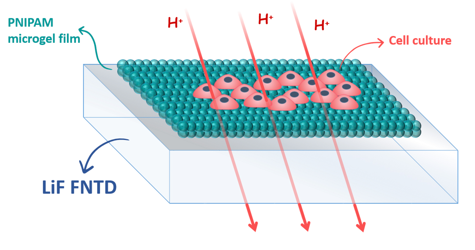
Figure 1 - Schematic of the prototype of the biocompatible detector obtained by combining the FNTD detector based on LiF and PNIPAM microgel film as a substrate for cell cultures
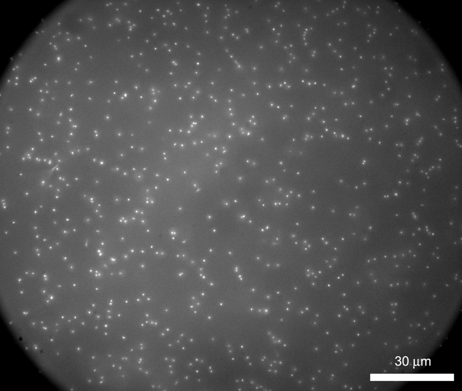
Figure 2 - Image obtained with a fluorescence microscope of single-proton traces from single protons with an energy of 1 MeV recorded in a LiF crystal
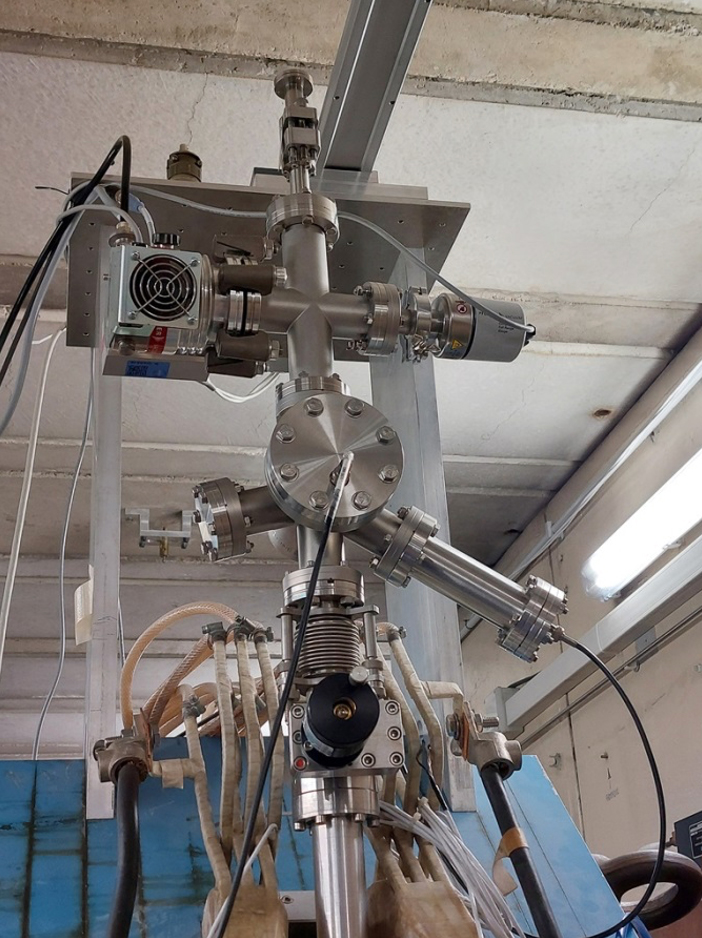
Figure3 - Photograph of the TOP-IMPLART vertical beamline
Notes
[1] Fluorescent Nuclear Track Detectors for radiobiology (FNTD): https://www.biotrack.enea.it/en
[2] Research Group Projects 2020 - POR FESR Lazio 2014-2020
[3] Radiobiology is the discipline that studies the effects of ionizing radiation on living matter and the mechanisms through which these effects occur.
[4] TOP-IMPLART has been developed by ENEA in collaboration with the Italian National Institute of Health (ISS) and the Regina Elena National Cancer Institute (IRE-IFO)
[5] V. Nigro, E. Buratti, F. Limosani, R. Angelini, F. Dinelli, S. Franco, E. Nichelatti, M. Piccinini, M. A. Vincenti, R. M. Montereali, B. Ruzicka, Spin-coating deposition of thermoresponsive microgel thin films, Colloids and Surfaces A: Physicochemical and Engineering Aspects, 674, 131918 (2023)